Leg Bones Diagram - The Complete List of Bodybuilding Leg Exercises and the ... : Learn vocabulary, terms and more with flashcards, games and other study tools.
Dapatkan link
Facebook
X
Pinterest
Email
Aplikasi Lainnya
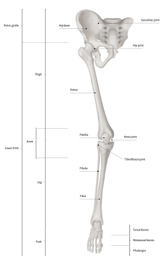
Leg Bones Diagram - The Complete List of Bodybuilding Leg Exercises and the ... : Learn vocabulary, terms and more with flashcards, games and other study tools.. Bones of the leg and foot, lower leg bone anatomy, leg bones anatomy, leg muscles, leg bones diagram, leg bone structure, leg anatomy muscles, parts of the lower leg. The patella in the knee; Normal leg bones are relatively straight, but those affected by paget's disease are porous and figure 9. Most bones (particularly the long bones of the arms and legs — which make up the appendicular skeleton) have a hard outer shell known as cortical bone. The foot bones shown in this diagram are the talus, navicular, cuneiform, cuboid.
Each leg is made up of four bones. Most bones (particularly the long bones of the arms and legs — which make up the appendicular skeleton) have a hard outer shell known as cortical bone. Visit kenhub for more skeletal system quizzes. The human leg, in the general word sense, is the entire lower limb of the human body, including the foot, thigh and even the hip or gluteal region. Master leg and knee anatomy using our topic page.
Arches of the foot: Anatomy | Kenhub from thumbor.kenhub.com Visit kenhub for more skeletal system quizzes. Human anatomy diagrams show internal. Bones of the leg and foot, lower leg bone anatomy, leg bones anatomy, leg muscles, leg bones diagram, leg bone structure, leg anatomy muscles, parts of the lower leg. The foot bones shown in this diagram are the talus, navicular, cuneiform, cuboid, metatarsals. Time to jump right into the biggest and strongest bones in the human body. The bones of the leg are the femur, tibia, fibula and patella. Your leg bones are the longest and strongest bones in your body. Click now to learn more about the bones, muscles, and soft tissues tibia:
Bones diagram human body 12 photos of the bones diagram human body all bones human body diagram, anatomy diagram human body.
Its lower end helps create the knee joint. Learn how to draw the femur, patella, tibia, and fibula in this lesson! The bones involved in it, however, are only the femur and the tibia, although the smaller bone of the leg, the fibula, is carried along in the movements of flexion, extension, and slight rotation that this joint. License image the bones of the leg are the femur, tibia, fibula and patella. Human anatomy diagrams show internal. Use the leg bones diagrams to learn the names of the leg bones. Normal leg bones are relatively straight, but those affected by paget's disease are porous and figure 9. High resolution textures and displacement included. However, the definition in human anatomy refers only to the section of the lower limb extending from the knee to. At the same time, the bones and joints of the leg and foot must be strong enough to support the body's weight while remaining flexible enough for movement and balance. Health diagram bone skeleton leg knee science anchor chart human human body. This bright worksheet helps your child bring these technical terms down to size. Click on the figures for a detailed view you will find the pelvic bones in the hip;
At the microscopic level, this hard outer. The bones of the leg are the femur, tibia, fibula and patella. The foot bones shown in this diagram are the talus, navicular, cuneiform, cuboid. Bones diagram human body 12 photos of the bones diagram human body all bones human body diagram, anatomy diagram human body. Visit kenhub for more skeletal system quizzes.
Toe Woes | Leg anatomy, Anatomy reference, Anatomy and ... from i.pinimg.com High quality realistic skeleton legs. At the same time, the bones and joints of the leg and foot must be strong enough to support the body's weight while remaining flexible enough for movement and balance. He'll boost his body knowledge as he matches up the names of the bones with their proper places on the leg diagram. Its lower end helps create the knee joint. Bones diagram human body 12 photos of the bones diagram human body all bones human body diagram, anatomy diagram human body. Bones of the leg and foot, lower leg bone anatomy, leg bones anatomy, leg muscles, leg bones diagram, leg bone structure, leg anatomy muscles, parts of the lower leg. Normal leg bones are relatively straight, but those affected by paget's disease are porous and figure 9. Blood vessels and nerves enter the bone.
High quality realistic skeleton legs.
The musculoskeletal segment of the leg, including the foot bones (ankle, heel bone, toe bones), fibula and tibia, knee, femur and femoral neck, hip and sacrum as well as the third, fourth. Human anatomy diagrams show internal. The bones involved in it, however, are only the femur and the tibia, although the smaller bone of the leg, the fibula, is carried along in the movements of flexion, extension, and slight rotation that this joint. Pngtree offers bone diagram png and vector images, as well as transparant background bone diagram clipart images and psd files. Quizzes on human skeletal system anatomy, bone anatomy, and bone markings. Bones of the leg and foot, lower leg bone anatomy, leg bones anatomy, leg muscles, leg bones diagram, leg bone structure, leg anatomy muscles, parts of the lower leg. Your leg bones are the longest and strongest bones in your body. Click now to learn more about the bones, muscles, and soft tissues tibia: The femur, or thighbone, is the longest and largest bone in the human body. Diagram of blood and nerve supply to bone. Normal leg bones are relatively straight, but those affected by paget's disease are porous and figure 9. Click on the figures for a detailed view you will find the pelvic bones in the hip; Time to jump right into the biggest and strongest bones in the human body.
At the same time, the bones and joints of the leg and foot must be strong enough to support the body's weight while remaining flexible enough for movement and balance. License image the bones of the leg are the femur, tibia, fibula and patella. The human leg consists of 8 bones, 4 per leg. Time to jump right into the biggest and strongest bones in the human body. This bright worksheet helps your child bring these technical terms down to size.
Infographic Diagram Of Human Skeleton Lower Limb Anatomy ... from media.istockphoto.com Visit kenhub for more skeletal system quizzes. The patella in the knee; The foot bones shown in this diagram are the talus, navicular, cuneiform, cuboid, metatarsals and calcaneus. Includes leg (femur, tibia, patella, and fibula) and foot (tarsals and digits) bones. Normal leg bones are relatively straight, but those affected by paget's disease are porous and figure 9. Pngtree offers bone diagram png and vector images, as well as transparant background bone diagram clipart images and psd files. High quality realistic skeleton legs. The human leg, in the general word sense, is the entire lower limb of the human body, including the foot, thigh and even the hip or gluteal region.
Diagram of blood and nerve supply to bone.
When you stand or walk, all the weight of your upper body rests on them. He'll boost his body knowledge as he matches up the names of the bones with their proper places on the leg diagram. Learn vocabulary, terms and more with flashcards, games and other study tools. Most bones (particularly the long bones of the arms and legs — which make up the appendicular skeleton) have a hard outer shell known as cortical bone. Quizzes on human skeletal system anatomy, bone anatomy, and bone markings. Click now to learn more about the bones, muscles, and soft tissues tibia: Your leg bones are the longest and strongest bones in your body. The foot bones shown in this diagram are the talus, navicular, cuneiform, cuboid, metatarsals. Pngtree offers bone diagram png and vector images, as well as transparant background bone diagram clipart images and psd files. The musculoskeletal segment of the leg, including the foot bones (ankle, heel bone, toe bones), fibula and tibia, knee, femur and femoral neck, hip and sacrum as well as the third, fourth. However, the definition in human anatomy refers only to the section of the lower limb extending from the knee to. Related posts of bones leg diagram picture. At the microscopic level, this hard outer.


Komentar
Posting Komentar